Assembling an accurate image of what the inside of an organ looks like is not an easy task. In order to figure out what the inside of a liver or eyeball or brain looks like, said organs often need to be sliced into tiny slivers, which are then individually studied with a microscope. But now, another method of studying the insides of organs—which utilizes AI and a process called “tissue clearing”—is allowing researchers to study these biological structures in a way that’s not only far less work-intensive, but also more conducive to understanding how they actually work.
1) We are super excited to present VesSAP with Bjoern Menze lab: a novel machine learning technology to segment complete mouse brain vasculature now online at @naturemethods https://t.co/VDAGnc0lbq
— Ali Max Erturk (@erturklab) March 11, 2020
work by @jocpae & @MihailMuc #AI #deeplearning #machinelearning #vessel #3D pic.twitter.com/JTUSEC7oQA
Futurism reported on the new process, which has been dubbed “Small-micelle-mediated Human orgAN Efficient clearing and Labeling” or SHANEL. The process is being developed by researchers in Germany, from Helmholtz Zentrum München (a research center), the LMU University Hospital Munich, and the Technical University of Munich. And in a recent paper by Ali Ertürk, et al., published in the journal, Nature, SHANEL was used to generate a transparent, 3D image of the whole vasculature of a mouse brain.
Ertürk, and a doctoral student studying under him who worked on the project, Mihail Ivilinov Todorov, spoke with Neuroscience News about the cutting-edge visualization, and highlighted the fact that having a clear picture of the entirety of a brain’s vasculature (the way its blood vessels are arranged, how large they are, etc.), helps us to better understand both its normal and pathological functions. Traumatic brain injuries, for example, cause perturbations in normal vascular structure and function in the brain, as do diseases like Alzheimer’s disease.
![]()
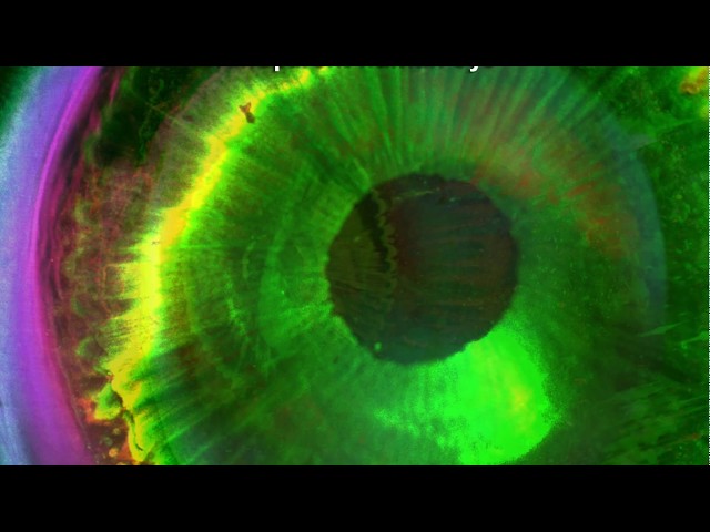
A video showing organs that have been imaged using SHANEL.
As mentioned, SHANEL works using a combination of tissue clearing and AI, and, thanks especially to the latter, is able to image organs like never before. While the process of tissue clearing—whereby an organ is made transparent through the use of special dyes that change the refractive index of its component tissues, and therefore allow them to uniformly absorb and reemit light—has been established for some time, Neuroscience News notes that Ertürk and his team took a new approach, and combined multiple dyes, which allowed them to see both the large and small vessels in their sample mouse brain simultaneously.

Mouse neurons imaged using SHANEL. Ertürk, et al.
Once the researchers had used their novel dye combination to make the mouse brain transparent, they then captured images of its vasculature, and handed those off to another team at the Technical University of Munich, led by Björn Menze, which used AI to assemble those images in an accurate and cohesive model. More specifically, Menze and his team used machine learning to accurately reconstruct the complete vasculature from the incomplete or skewed images taken of the transparent mouse brain.
“Over the past few years, we have developed a deep learning algorithm that specialises in detecting blood vessels in medical images,” Menze told Neuroscience News, adding that “This was the first time we applied it to a whole brain.” The algorithm developed by Menze and his team is able to reliably identify blood vessels from other tissues, even if original images are not well-illuminated or distorted due to other errors in source images. (SHANEL has also been used previously to generate transparent 3D images of other organs, including a human eye, a human brain, and a human kidney, although their vasculature was not imaged with this kind of clarity.)
We are very excited to introduce SHANEL to map adult human organs. It employs a new tissue permeabilization chemistry for labeling and clearing.
— Ali Max Erturk (@erturklab) May 21, 2019
We generated the first transparent adult human brain.
Developed by amazing scientists @shan_heather et al.https://t.co/kgnf0DsKk7 pic.twitter.com/xkAQlNEwLQ
These mouse-related findings are, of course, only the beginning of the story for SHANEL. “With our system, we are likely to be able to analyse the small tissue specimens from human [tumors] with greater accuracy,” Ertürk said, adding that this could have “an optimizing effect” on treatment. According to Neuroscience News, the researchers also want to eventually use the visualizations developed using SHANEL to create a 3D printer that can crank out usable human organs.
What do you think about SHANEL and its ability to generate astoundingly detailed images of organs? And how soon do you think we’ll be 3D printing livers, hearts, and eyeballs? Let us know your thoughts in the comments!
Feature image: Mihail Ivilinov Todorov